
5 Conditions a Professional Optometrist Can Detect
Maintaining clear sight and vibrant ocular health requires routine evaluation by a qualified specialist. Many individuals assume that visual examinations are only intended for updating visual prescriptions or obtaining new corrective lenses. However, a comprehensive assessment provides a deep look into the structural health of your eyes and can uncover early indicators of various systemic and localized health issues. Because many serious ocular conditions develop silently without initial pain, discomfort, or obvious warning signs, booking routine visual evaluations plays a critical role in proactive personal health management. Establishing a routine schedule for professional evaluations allows you to stay informed about subtle changes occurring within your visual system over time.
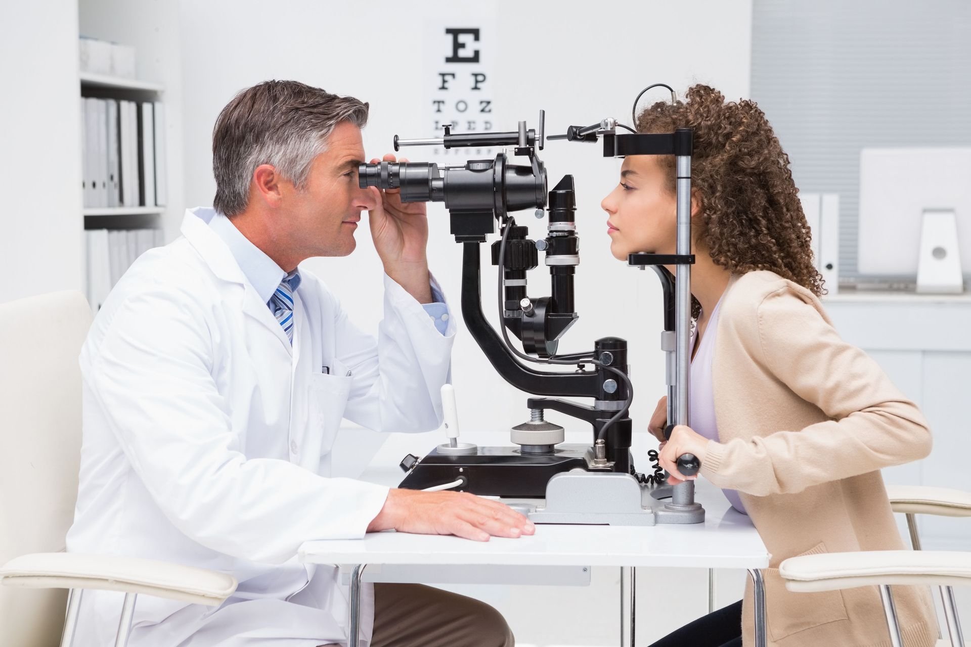
When you schedule a thorough evaluation, an optometrist utilizes specialized visual assessment tools and advanced light sources to inspect both the interior and exterior structures of the eye. This comprehensive analytical process allows for the early identification of potential physiological concerns long before they begin to cause noticeable visual impairment or permanent damage. Detecting subtle structural changes early opens up a wider array of management options and helps preserve long-term visual clarity and quality of life.
1. Glaucoma
Glaucoma is often referred to as a silent threat to visual wellness because it typically progresses without early symptoms, pain, or visible visual changes. This progressive ocular condition involves gradual degradation of the delicate optic nerve fibers, which are responsible for transmitting visual signals directly from the eye to the visual cortex of the brain. In many cases, elevated fluid pressure within the anterior chamber of the eye contributes to this neural breakdown, though normal-tension variations also occur frequently across diverse populations. Understanding how fluid dynamics operate within the globe helps illustrate why regular monitoring remains indispensable for individuals of all backgrounds.
Without consistent routine monitoring, severe peripheral vision loss can occur before an individual even realizes that functional visual field changes are taking place. During a routine physical assessment, an optometrist measures fluid pressure levels within the interior chamber and examines the precise structural appearance of the optic nerve head using high-magnification tools. Advanced diagnostic instruments allow for the measurement of subtle changes in nerve fiber layer thickness long before functional vision loss manifests in daily activities. Early detection remains vital because structural damage caused by nerve fiber degradation cannot be reversed, but timely management strategies can effectively slow down or halt further functional progression.
2. Cataracts
Cataracts represent a natural progressive clouding of the crystalline lens inside the eye, which normally functions to focus light precisely onto the sensitive retinal tissue located at the back of the globe. Over time, delicate proteins within the lens structure begin to clump together, creating hazy, blurred, distorted, or muted visual perception. Individuals experiencing this common anatomical issue often report feeling as though they are looking through a perpetually foggy or dirty window. Other common secondary indicators include heightened sensitivity to bright light sources, persistent glare while navigating at night, and reduced contrast when viewing subtle color variations.
Through specialized high-magnification equipment, an optometrist evaluates the overall density, location, and structural clarity of the natural lens. Identifying these localized tissue changes in their initial stages allows for proper tracking over extended periods. While early stages might simply require simple environmental modifications such as improved task lighting or updated lens prescriptions, detailed ongoing monitoring ensures that appropriate medical steps are recommended whenever visual clarity begins to interfere with daily activities or personal safety. Consistent monitoring allows individuals to make informed decisions regarding their ongoing visual comfort.
3. Diabetic Retinopathy
Systemic metabolic conditions can have a direct and profound impact on microvascular networks throughout the body, particularly within the sensitive layers of the retinal tissue. Diabetic retinopathy occurs when elevated blood glucose levels cause delicate capillary blood vessels in the back of the eye to swell, leak fluid, or become blocked entirely over time. In more advanced cases, fragile new blood vessels grow abnormally across the surface of the retina, leading to severe visual disruption, scar tissue formation, or total retinal detachment if left unmanaged.
A detailed internal evaluation allows an optometrist to visualize the microscopic blood vessels of the retinal landscape directly without invasive surgical procedures. Early physical indicators such as tiny microaneurysms, localized fluid accumulation, subtle hemorrhages, or vascular swelling can be detected during a standard appointment, often before a patient even receives an official systemic diagnosis from a primary care physician. Timely tracking of these microvascular changes helps coordinate appropriate collaborative care and protects long-term retinal integrity. Early intervention remains crucial for preventing permanent structural changes to the retina.
4. Macular Degeneration
The macula is the specialized central region of the retina responsible for sharp, detailed, straight-ahead vision necessary for crucial everyday tasks such as reading fine print, operating motor vehicles, recognizing facial features, and performing detailed work. Age-related macular degeneration involves the progressive breakdown of this critical retinal region, leading to persistent blurring, dark spots, or distorted perception of straight lines in the center of the visual field. While peripheral vision typically remains intact, the deterioration of central sight can significantly alter daily functionality and personal independence.
By conducting a detailed microscopic examination of the underlying retinal layers, an optometrist can identify tiny cellular waste deposits called drusen that gradually accumulate beneath the central retina. Observing these subtle structural shifts enables early lifestyle recommendations, targeted dietary adjustments, and structured ongoing observation strategies designed to safeguard central clarity and slow down the rate of retinal tissue degradation. Comprehensive care plans can be tailored to individual needs to maintain baseline functionality.
5. Color Vision Deficiency
Color vision deficiency, commonly referred to as color blindness, affects an individual's capacity to distinguish specific shades, hues, or color intensities, most frequently between red and green tones. This functional condition usually stems from genetic variations in the sensitive photopigments located within specialized cone photoreceptor cells in the central retina. While many individuals adapt well to living with altered color perception, identifying the precise nature of the issue early in life proves immensely helpful for educational environments, daily activities, and specific career path considerations.
According to the Cleveland Clinic, around 300 million people worldwide experience some form of color blindness. Specialized testing plates during a visual examination easily evaluate how accurately a person perceives subtle color variations and contrasts. An optometrist can administer these standardized visual assessments to determine the exact type and extent of the deficiency, offering helpful guidance on adaptive visual strategies for school, professional tasks, and everyday navigation. Learning about these variations empowers individuals to navigate daily environments with greater clarity and confidence.
Taking a proactive, consistent approach to personal ocular health is one of the most effective ways to safeguard your sight and maintain your quality of life for years to come. Routine comprehensive evaluations serve as a crucial line of defense, enabling early identification of asymptomatic visual conditions and supporting overall physical well-being. Prioritizing consistent professional care ensures that subtle changes within your ocular structures are identified, monitored, and addressed long before they affect your daily routine or long-term vision. Taking small proactive steps today protects your clear vision for all of tomorrow's experiences.
If you are ready for comprehensive visual care that prioritizes your long-term wellness, contact us at Jeffries Eye Care today. An optometrist on our team will help you maintain clear vision and protect your overall ocular health through personalized evaluations.







Share On: